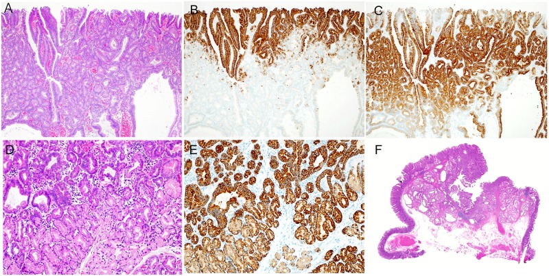
Fig 5. Histologic characteristics of Pyloric Gland-type (PG) belonging to gastric-type.
(A) The surface layer of a tumor (HE staining), showing hyperplasia of small atypical ducts and dilated atypical ducts. (B) The surface layer of a tumor (MUC5AC antibody staining), showing the presence of MUC5AC-positive cells in the superficial layer of the tumor. (C) The surface layer of a tumor (MUC6 antibody staining), showing the presence of MUC6-positive cells in a large area, excluding the superficial layer of the tumor. (D) The deep region of a tumor (HE staining), showing hyperplasia of small atypical ducts. A region showing hyperplasia of clear cells with no atypia was found in the deepest part of the tumor. (E) A deep part of a tumor (MUC6 antibody staining), showing the presence of MUC6-positive cells in a large area extending from the atypical ducts to the region of clear-cell hyperplasia. (F) A magnified image of a tumor (HE staining), showing a distinctly protruding lesion. A region of tumor duct and mucous duct hyperplasia was found in the lamina propria. The muscularis mucosa was compressed and pushed down deeply.

