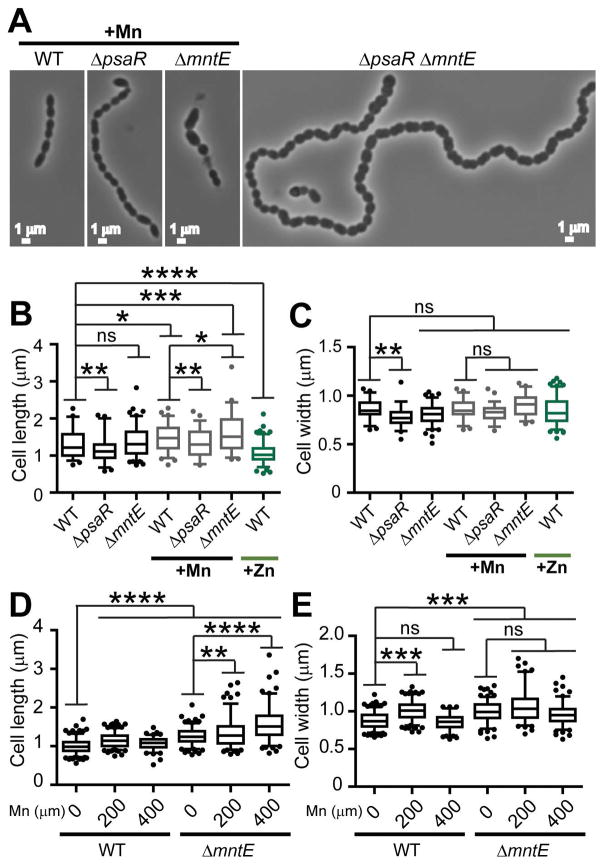
Figure 3. Cell division is disrupted by Mn-toxicity.
(A) Representative morphology of cells grown in BHI supplemented with 200 μM Mn at 3.5 h post-inoculation. No Mn was added to the double ΔpsaR ΔmntE mutant. Length (B) and width (C) measurements of cells grown with 0, 200 μM Mn, or 200 μM Zn. Length (D) and width (E) measurements of cells grown anaerobically with 0, 200, or 400 μM Mn. *P ≤ 0.10; **P ≤ 0.05; ***P ≤ 0.01; ****P ≤ 0.001; ns, not significant.

