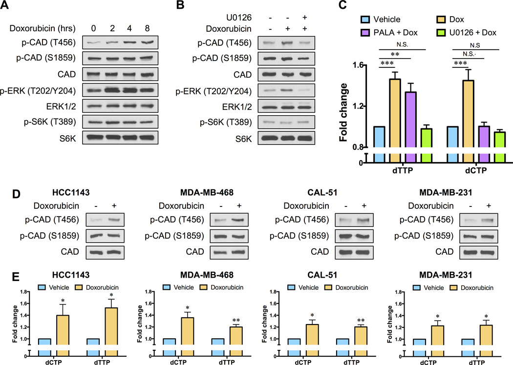
Fig. 2. Chemotherapy exposure alters the phosphorylation state of CAD to stimulate de novo pyrimidine nucleotide synthesis.
(A) SUM-159PT cells were treated with 0.5 µM doxorubicin for the indicated times and the phosphorylation states of CAD, ERK and S6K1 were monitored by immunoblotting. (B) SUM-159PT cells were pre-treated with 5 µM U0126 for 12 hours before a 4 hour exposure to 0.5 µM doxorubicin and the phosphorylation states of CAD, ERK and S6K1 were monitored by immunoblotting. (C) SUM-159PT cells were treated with 0.5 µM doxorubicin for ten hours, in the absence or presence of 5 µM U0126 or 40 µM N-(phosphonacetyl)-l-aspartic acid (PALA), and pyrimidine deoxyribonucleoside triphosphate levels were monitored using a fluorescence-based assay. (D) TNBC cell lines (HCC1143, MDA-MB-468, CAL-51 and MDA-MB-231) were treated with 0.5 µM doxorubicin for 4 hours and changes in CAD phosphorylation were monitored by immunoblotting. (E) TNBC cell lines (HCC1143, MDA-MB-468, CAL51 and MDA-MB-231) were treated with 0.5 µM doxorubicin for 10 hours and pyrimidine deoxyribonucleoside triphosphate levels were monitored using a fluorescence-based assay. All error bars represent SEM. N.S. not significant, * P < 0.05, ** P < 0.01, *** P < 0.001 by a Student’s t-test.

