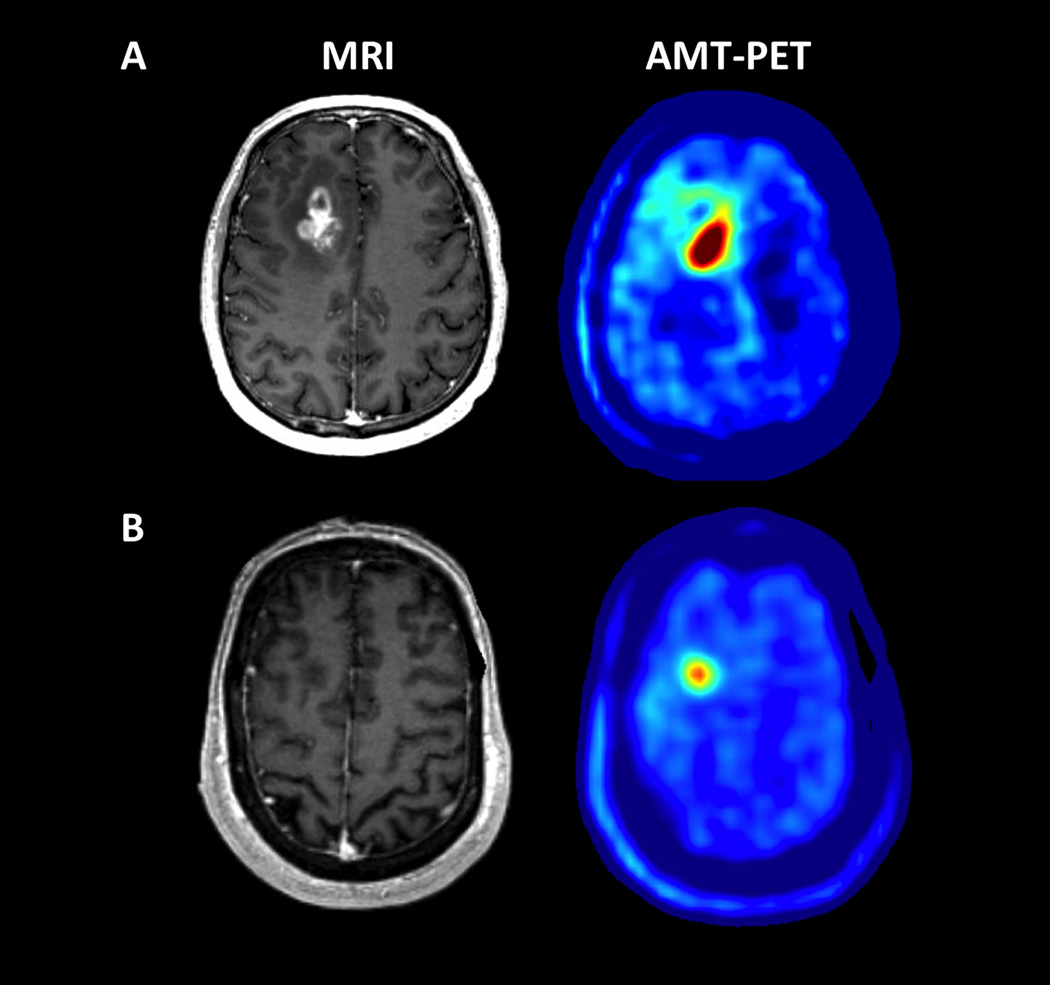
Figure 1.
Representative examples of AMT-PET in a patient with high (A) vs. low (B) uptake associated with different survival. (A) A 54 year-old male (patient #8 in Table 1) with a right frontal glioblastoma, measured with a 2.59 tumor/cortex AMT SUV-ratio, above the ROC-defined threshold of 1.94. He survived for more than 2 years after the PET scan. (B) A 68 year-old male (patient #10 in Table 1) with a right frontal glioblastoma showing a below-threshold tumor/cortex AMT SUV-ratio (1.56). He survived for only 7 months.

