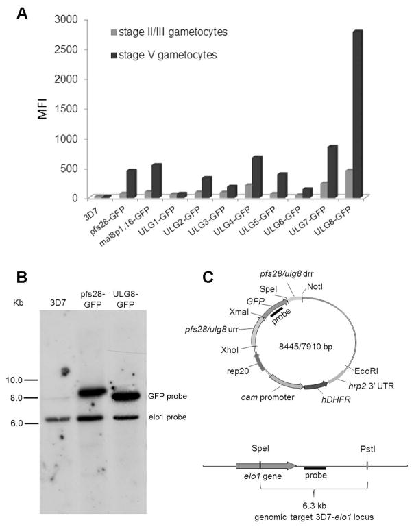
Figure 1. Development of the P. falciparum line 3D7/pfULG8-GFP upregulating GFP expression in stage V gametocytes.
A: Histograms representing the mean fluorescent intensity (MFI) of the GFP reporter expressed under control of the pfULG 1–8, the pfs28 and the mal8p1.16 regulatory regions in stage II/III and in stage V gametocytes (representative of two biological replicates). B. Southern blot analysis of genomic DNA from lines 3D7wt, 3D7/pfULG8-GFP and 3D7/pfs28-GFP. Equal amounts of SpeI+PstI digested genomic DNAs from the indicated parasite lines were electrophoresed and hybridized with probes specific for the GFP coding sequence and the single copy gene pfelo1 (see panel C). Autoradiographs from the two hybridizations were combined in the panel and hybridization bands specific for each probe are indicated. C. Diagram of the plasmid containing the GFP coding sequence flanked by the pfs28 or the pfULG8 regulatory regions (above) and of the chromosomal locus of the single copy gene pfelo1 (below) are shown to indicate position of the GFP and the pfelo1 specific probes, as are the positions of the SpeI and the PstI restriction sites used in the hybridization experiments in panel B. urr: upstream regulatory region; drr: downstream regulatory region.

