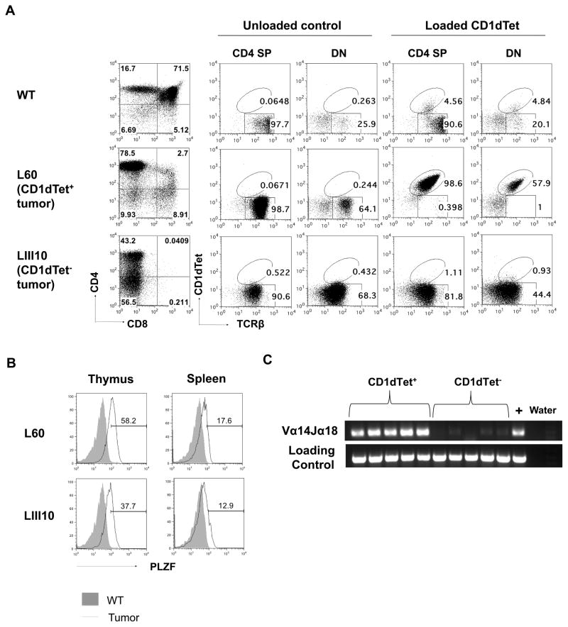
Figure 2. Lymphomas in L-DKO mice are CD1dTet+ (iNKT) or CD1dTet− in origin.
(A) Representative staining of thymocytes with CD4 and CD8 markers from a Cre− control and two L-DKO mice with either CD1dTet+ (L60) or CD1dTet− (LIII10) tumor. CD4 and DN fractions were further analyzed by staining with TCRβ and CD1dTet without or with loaded antigen. (B) Representative histogram of intracellular PLZF staining of TCRβ+ populations from mice (N = 2) with tumor shown in (A). (C) Detection of Vα14Jα18 rearrangement in CD1dTet+ or CD1dTet− lymphoma samples by PCR. CD14 was used as a loading control. (N = 5)

