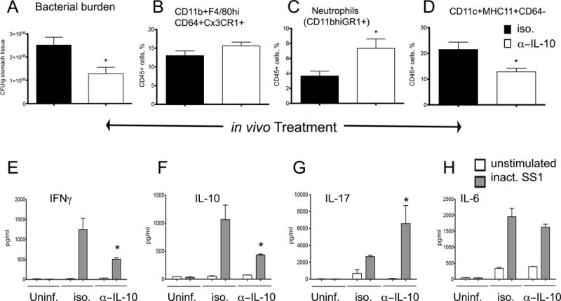
Figure 5. IL-10 neutralization during Helicobacter pylori infection.

WT mice were infected with H. pylori and on days 17, 19 and 21 post-infection they were treated with 100 μg of either neutralizing anti-mouse IL-10 or Rat IgG1 isotype control antibodies. Mice (n=6), were euthanized on day 22 post-infection to measure (A) bacterial burden in the stomach, and percentages of (B) CD11b+F4/80hiCD64+CX3CR1+, (C) neutrophils, and (D) DC. Cells from the gastric lymph nodes of 3–4 mice of the same gender and treatment were stimulated ex-vivo with RPMI (unstimulated) or 5 μg/ml of formalin-inactivated H. pylori SS1. Production of (E) IFNγ, (F) IL-10, (G) IL-17 and (H) IL-6 were measured in 72-hour cell culture supernatant using a cytometric bead array. Results are expressed as average of 3–5 samples of pooled cells and data represent mean ± SEM. Points with an asterisk are significantly different when compared to the control group (P<0.05). Results are representative of 2 independent replicate experiments with same results. Points with an asterisk are significantly different when compared to the control (WT) group (P<0.05).
