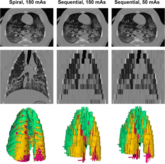
Fig. 2.

Scanning protocols. Axial (upper panels), coronal (middle panels) and 3D reconstructions (lower panels) of the three acquisition protocols in a representative animal. Colors in the 3D reconstruction represent clusters of lung tissue: hyper-aerated (blue), normally aerated (green), poorly aerated (yellow), non-aerated (red)
