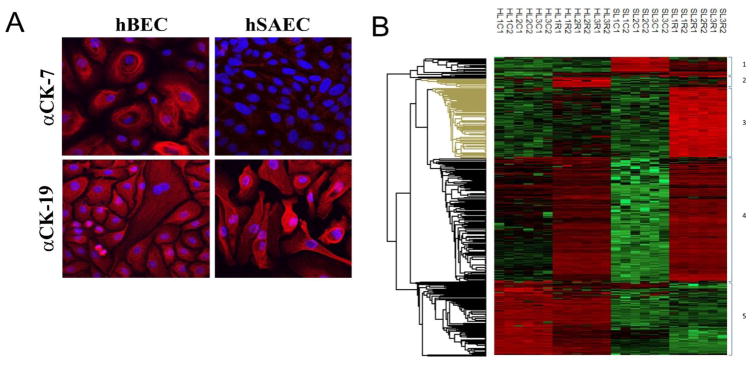
FIGURE 5.
Identification of differentially expressed proteins in primary airway epithelial cells. (A). Immunofluorescence of differential cytokeratin expression. phBECs or phSAECs plated on collagen coverslips were fixed and stained with anti-cytokeratin (CD)-7 or 19-selective Abs. Secondary detection was Alexa Fluor 568 (red)-conjugated goat anti-rabbit IgG. Nuclei were counterstained with DAPI and images acquired by confocal microscopy. For each antigen, the merged DAPI image is shown. (B). Hierarchical clustering of secretome proteins from primary airway cells. Differentially expressed proteins identified in the secretomes of primary phBECs and primary phSAECs through the analysis in Figs. 2–4 are shown. Log2-transformed expression values for biological replicates values were z-score-normalized and subjected to hierarchical clustering. Shown are individual data for each biological replicate. Abbreviations used are: H, phBECs; S, phSAECs; L#, lot number (different donors); C, control; R, RSV; Arabic numbering (1,2) indicates technical replicate.

