
Fig. 4.
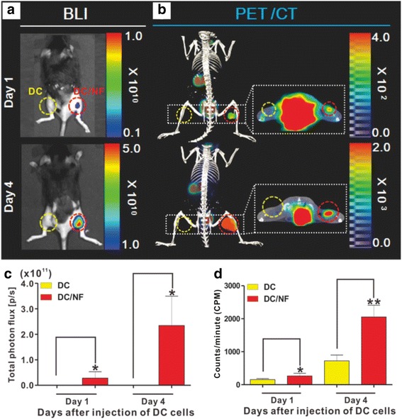
In vivo 18F-TFB PET/CT imaging and BLI of intramuscularly injected DCs. Parental DC and DC/NF cells were intramuscularly injected into the left and right thighs of mice, respectively, and combined 18F-TFB PET/CT imaging and BLI was performed. a In vivo BLI and b 18F-TFB PET/CT imaging of parental DC and DC/NF cells on days 1 and 4 after the intramuscular injection. The red and yellow circles indicate the DC and DC/NF injection sites, respectively. ROI analysis of c bioluminescent and d PET/CT images. Data are expressed as mean ± SD; n = 5