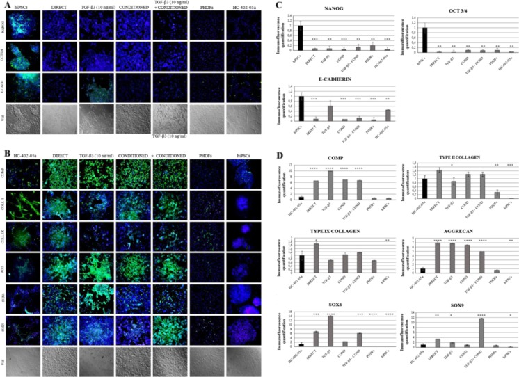
Fig. 3.
The presence of chondrogenic markers was demonstrated by immunofluorescence analysis. The human-induced pluripotent stem cells (hiPSC)-derived chondrocytes: DIRECT, TGF-β3, COND, TGF-β3+ COND did not reveal the presence of pluripotency markers: Nanog, OCT4, E-cadherin (a), but they did express desirable chondrogenic proteins: COMP, type II and IX collagen, aggreccan (AGG), SOX6 and SOX9 (b). The pluripotency and chondrogenic markers were quantified to better show the differences between particular differentiation protocols (c and d)

