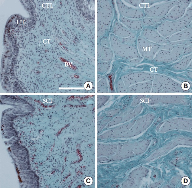
Fig. 2.

(A) Comparison of the urinary bladder wall of a selected healthy reference minipigs (CTL) to a selected spinal cord injury (SCI) minipig. (A, C) Urothelial and suburothelial region of the urinary bladder wall. (B, D) Detrusor smooth muscle region of the urinary bladder wall. BV, blood vessel; CT, connective tissue; MT, smooth muscle tissue; UT, urothelial tissue. Masson-Goldner staining. The scale bars represent 1 mm.
