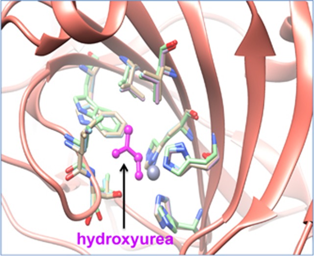
Figure 7.
Superposition of X-ray crystal structures of carbonic anhydrase 2 (CA2) and its paralogs (CA13, CA7) bound to hydroxyurea. The ribbon structure of CA2 and its paralogs is shown in pink color. The hydroxyurea in the protein pocket is shown in magenta color bound to amino acid residues that are conserved across CA2 and its paralogs.

