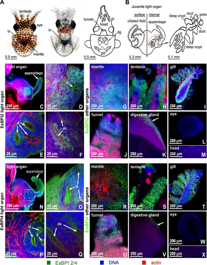
FIG 4 .
Localization of EsBPI2 and EsBPI4 in juvenile squid tissues. Proteins were probed with rabbit anti-EsBPI2 or anti-EsBPI4 antibodies, which were detected with FITC-conjugated goat anti-rabbit secondary antibody (EsBPI2/4 [green]), and counterstained with rhodamine-phalloidin (actin cytoskeleton [red]) and TOTO-3 (blue) (nuclei). (A) Dorsal and ventral views of the juvenile squid, and diagram to illustrate internal regions depicted in the following panels of the figure. In the diagram, the digestive gland (dg) is shown. lo, light organ. (B) Enlargement of the light organ showing the surface in contact with the environment (left) and the internal structure (right) where symbionts colonize. (Right) Anatomical structures of the three crypts indicated by the dashed red circle. (C to F) EsBPI2 ICC labeling at the top surface of the light organ (C), in the pore (p) (white arrow) (D), in the ducts (d) (white arrows) (area of white dashed box in panel C) (E), and in the deep crypts (dc) (deep crypt lumen indicated by white arrows) (white dashed circle in panel C, which is tissue superficial to the deep crypts) (F). (G to M) Localization of EsBPI2 ICC labeling in other organs examined. The white arrow points to the epithelial edge of the digestive gland. (N to Q) EsBPI4 ICC labeling on the top surface of the light organ (N), in the pores (p) (white arrows) (O), in the ducts (d) (white arrows) (white dashed box area in panel N) (P), and in the deep crypts (dc) (white arrows indicate deep crypt lumen) (white dashed circle in panel N, which is tissue superficial to the deep crypts) (Q). (R to X) Localization of EsBPI4 ICC labeling in other organs examined. The white arrow points to the epithelial edge of the digestive gland.

