
FIG 7 .
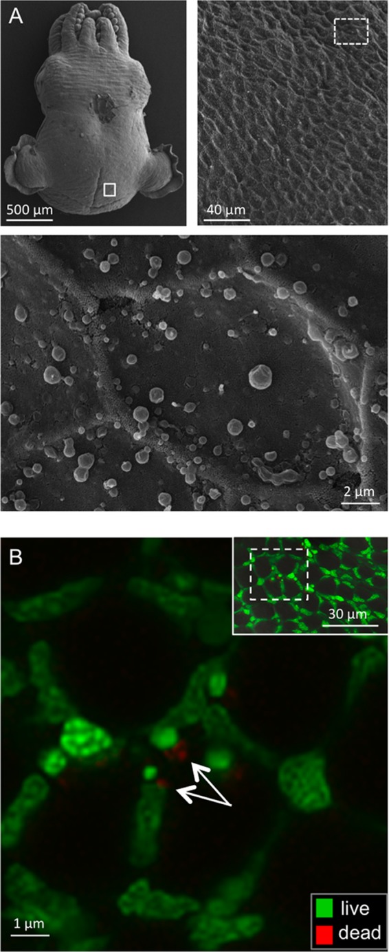
Detection of microbes associating with the outer mantle surface of E. scolopes. (A) Scanning electron microscopy (SEM). (Top, right and left) Low-magnification images of the animal showing the region examined for microbes (white boxes). (Bottom) High-magnification image of the epithelial surface (n = 4) (the whole skin of the animal was examined) with no microbes detected. (The irregular, round globules on the surface are mucous secretions from the skin.) (B) Confocal micrographs of a live, anesthetized animal that had been washed with filter-sterilized seawater three times for 5 min each time and then labeled for detection of live (green) or dead (red) bacterial cells. Images were taken in the same surface region as for SEM. (Inset, top right) Low-magnification view to show region (box outlined with white dashed line) examined at higher magnification. White arrows indicate possible vestiges of dead bacterial cells (n = 5).