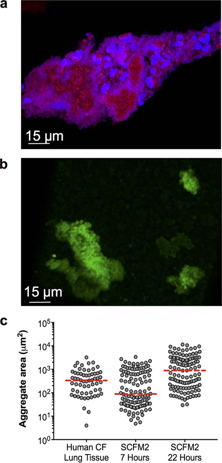
FIG 1 .

P. aeruginosa aggregate sizes are similar in human CF tissue and SCFM2. (a) Micrograph of a P. aeruginosa aggregate in an explanted human CF lung tissue section. P. aeruginosa was labeled by FISH. P. aeruginosa cells are red, and polymorphonuclear leukocytes are blue. (b) Confocal micrograph of a GFP-expressing P. aeruginosa aggregate in SCFM2 at 22 h postinoculation. (c) Cross-sectional area of P. aeruginosa aggregates from explanted CF lung tissue and P. aeruginosa aggregates grown for 7 and 22 h in SCFM2. Each symbol represents an aggregate, and the median is indicated by a broken red line. Measurements were accomplished by manually outlining aggregates, and the area within the outline was calculated with Fiji image analysis software. For these measurements, aggregates were assumed to be entirely filled with cells.
