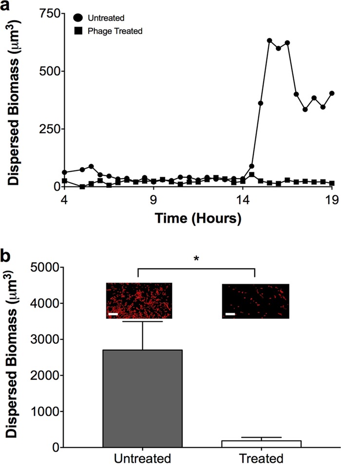
FIG 5 .

Phage reduce bacterial migrant numbers and prevent P. aeruginosa dispersal. (a) Dispersed P. aeruginosa biomasses during growth in SCFM2 in the presence (■) and absence (●) of phage. P. aeruginosa was allowed to form aggregates for 4 h in SCFM2 before phage addition, and the dispersed biomass was assessed every 30 min for 15 h. Dispersed biomass was defined as individual cell clusters of ≤5.0 µm3, which corresponds to about one to five bacteria. Data from a single replicate are presented for clarity. Three additional replicates are shown in Fig. S7. (b) Dispersal of P. aeruginosa in SCFM2. P. aeruginosa was allowed to form aggregates for 4 h in SCFM2 and then overlaid with sterile SCFM2 (untreated) or SCFM2 containing phage (treated). After 6 h, the amount of dispersed biomass in the upper SCFM2 layer was determined as the number of red fluorescent voxels and is represented as the mean biomass ± the standard error of the mean of four replicates. Representative micrographs of dispersed biomass are shown above each bar. Scale bar is 20 µm. An asterisk denotes a statistically significant difference between untreated and treated samples, as determined by a two-tailed t test [t(6) = 2.5, P = 0.044].
