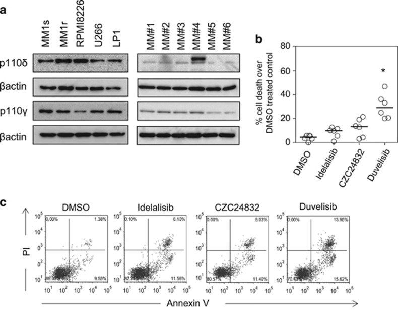
Figure 1.
Inhibition of PI3Kδ and PI3Kγ subunits in MM proliferation. (a) Western blot analysis of PI3Kδ and PI3Kγ subunits in MM cell lines and MM primary cells. Blots were probed with β-actin to show sample loading. (b) MM primary cells were treated with idelalisib (1 μm), CZC24832 (1 μm) and duvelisib (1 μm) for 72 h and then assessed for cell viability using CellTiter Glo. (c) MM primary cells were treated with idelalisib (1 μm), CZC24832 (1 μm) and duvelisib (1 μm) for 72 h and then assessed for apoptosis by PI/Annexin V staining (n=4).

