Figure 3.
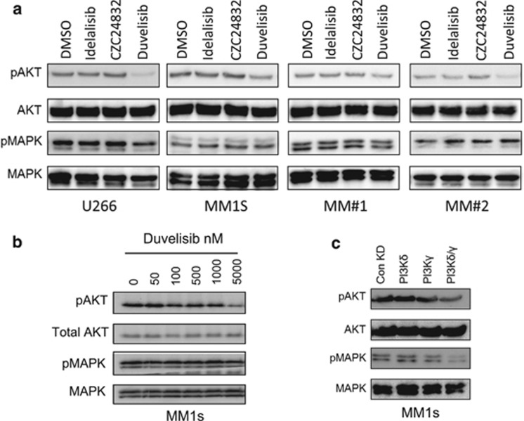
Inhibition of PI3Kδ and PI3Kγ inhibits AKT phosphorylation. (a) MM1s, U266, MM#1 and MM#2 cells were incubated with idelalisib (1 μm), CZC24832 (1 μm) and duvelisib (1 μm) for 4 h after which protein was extracted. Samples were subsequently analysed for phospho-AKT and phospho-MAPK responses using Western blotting. Blots were re-probed for total AKT and MAPK to confirm sample loading. (b) MM1s were incubated with increasing doses of duvelisib for 4 h after which protein was extracted. Samples were then analysed for phospho-AKT and phospho-MAPK response using Western blotting. Blots were re-probed for total AKT and MAPK to confirm sample loading. (c) MM1s cells were transduced with lentivirus targeted to PI3Kδ and PI3Kγ or control shRNA for 72 h. Protein was extracted and analysed for phospho-AKT and phospho-MAPK via Western blotting. Total AKT and MAPK were used as loading controls.

