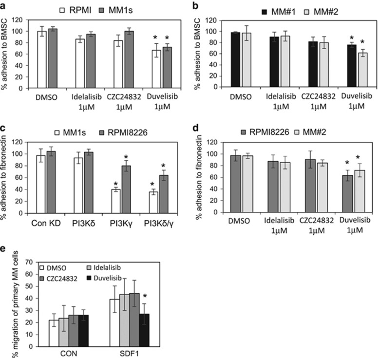
Figure 4.
MM adhesion to BMSC and fibronectin is regulated by PI3Kδ. (a, b) MM cell lines and MM primary cells were treated with idelalisib (1 μm), CZC24832 (1 μm) and duvelisib (1 μm) for 4 h and then stained with calcein AM. Stained cells were added to BMSC on 96-well plates for a 4 h incubation. Non-adherent cells were removed and fluorescence was measured. (c) MM1s and RPMI8226 cells were transduced with lentivirus targeted to PI3Kδ and PI3Kγ or control shRNA for 72 h. Transduced cells were stained with calcein AM and added to BMSC on a 96-well plate for a 4 h incubation. Non-adherent cells were removed and fluorescence was measured. (d) RPMI8226 and MM#2 primary cells were treated with idelalisib (1 μm), CZC24832 (1 μm) and duvelisib (1 μm) for 4 h and then stained with calcein AM. Stained cells were added 96-well plates coated with fibronectin for a 4 h incubation. Non-adherent cells were removed and fluorescence was measured. (e) MM cell lines were incubated with idelalisib (1 μm), CZC24832 (1 μm) and duvelisib (1 μm) for 4 h and subsequently treated with calcein AM. Cells were then placed in the upper well of a 5 μm transwell plate. The lower chamber contained 30 μl of serum-free media or serum-free media supplemented with SDF1 (100 ng/ml) for 4 h and MM cell migration was then assessed using fluorescent plate reader.

