Figure 1.
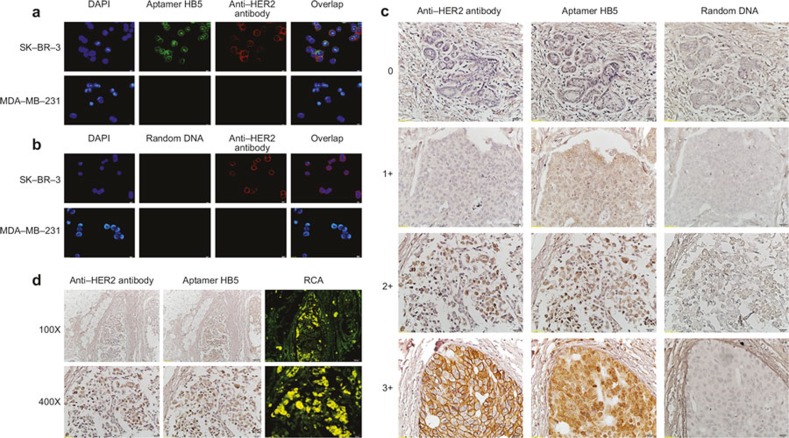
Figure 1. Evaluation of HER2 in breast cancer with the aptamer HB5. (a) Binding profiles of the aptamer HB5 to HER2-positive (SKBR3) or HER2-negative (MDA-MB-231) breast cancer cells. (b) The binding profiles of random DNA to breast cancer cells. (c) Examples of IHC staining for HER2 in invasive ductal carcinoma with a commercial anti-HER2 antibody or the aptamer HB5. (d) The detection of HER2 expression in situ based on the RCA.

