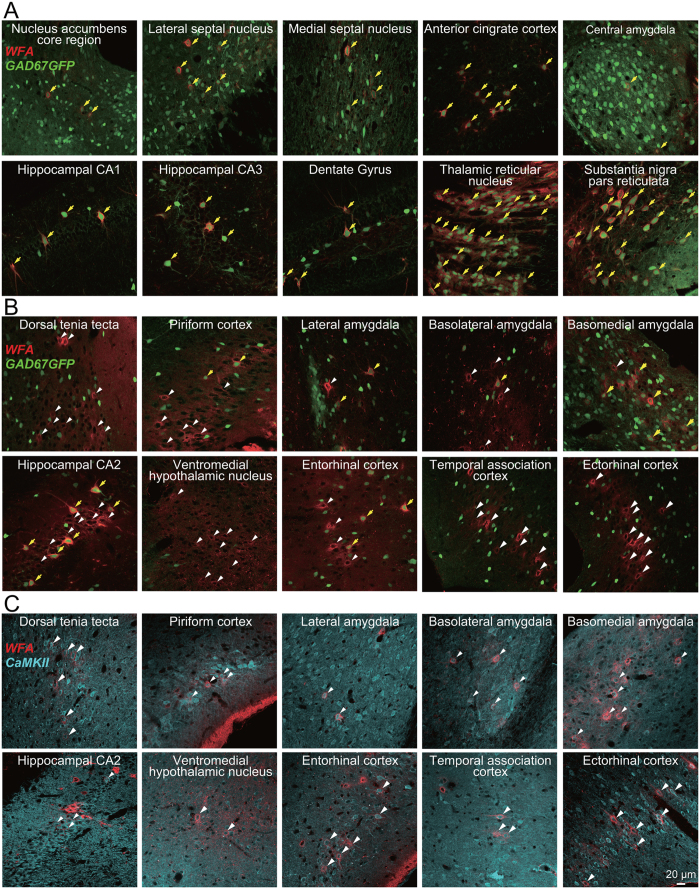
Figure 1. Distribution of PNNs throughout the whole mouse brain.
Merged images showing signals generated by WFA (red), GFP (green), or an anti-CaMKII antibody (cyan) in GAD67-GFP knock-in mice (A,B) or C57BL/6J mice (C). A subpopulation of GFP-expressing GABAergic neurons is enwrapped by WFA-positive PNNs (yellow arrows; A,B); however, WFA-positive PNNs also enwrap some neurons that do not express GFP (white arrowheads; B) or neurons that express CaMKII (white arrowheads; C).

