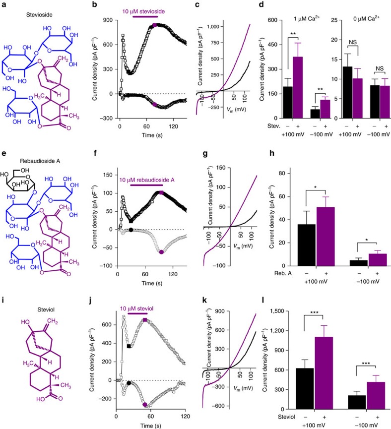
Figure 1. TRPM5-mediated currents are potentiated by stevioside and rebaudioside A and their aglycon steviol.
(a) Structure of stevioside. (b) Time course of inward and outward currents from TRPM5 overexpressing HEK cells. To activate TRPM5, pipette solution contained 1 μM free Ca2+. Time 0 indicates break-in into the cell. Stevioside was applied as indicated. (c) Representative current traces from the time points indicated in b. (d) Left: the average±s.e.m. (n=13 cells, paired t-test, P<0.01) peak inward and outward current before and during the application of stevioside with 1 μM intracellular Ca2+. Right: the average±s.e.m. inward and outward current before and during the application of stevioside in the absence of free intracellular Ca2+ (n=10 cells, P>0.2, paired t-test). (e) Structure of rebaudioside A. (f) A time trace with inward and outward currents from Trpm5 overexpressing HEK cells. Rebaudioside A was applied as indicated. (g) Current traces upon the application of rebaudioside A as indicated in f. (h) Average±s.e.m. (n=14 cells, paired t-test P<0.05) inward and outward current before and during application of rebaudioside A. (i) Structure of steviol. (j) A time trace with inward and outward currents from Trpm5 overexpressing HEK cells. Steviol was applied as indicated. (k) Current traces upon the application of steviol as indicated in j. (l) Average±s.e.m. (n=12 cells, paired t-test P<0.001) inward and outward current before and during application of steviol. See also Supplementary Figs 1 and 2. Reb. A, rebaudioside A; Stev., stevioside; NS, not significant.

