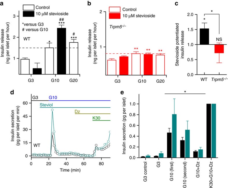
Figure 4. Stevioside potentiates glucose-induced insulin secretion in vitro and in vivo.
(a) Insulin release measured in vitro on individual isolated islets of WT and (b) Trpm5−/− mice (average±s.e.m. from n=24–30 islets per condition from four mice per genotype, two-sample t-test). The horizontal dashed line represents the level of the (10 mM) GIIS. (c) The effect of stevioside administration on glucose-induced insulin release in plasma from eight WT and nine Trpm5−/− mice (average±s.e.m). (d) Example trace of insulin secretion during perifusion experiments without steviol (black) or with steviol supplementation at 10 min in 3 mM glucose (G3), 10 mM glucose (G10) and with 100 μM diazoxide (Dz) and 30 mM KCl (K30). (e) Total insulin secretion in different conditions relative to the K30 stimulus. First GIIS is the initial insulin peak between 20 and 30 min in c and the second phase between 30 and 50 min. Significant difference between control (n=6 experiments) and steviol conditions (n=6) of 866 islets from 22 WT mice with a two-way analysis of variance. See also Supplementary Fig. 5 and Supplementary Table 1. NS, not significant.

