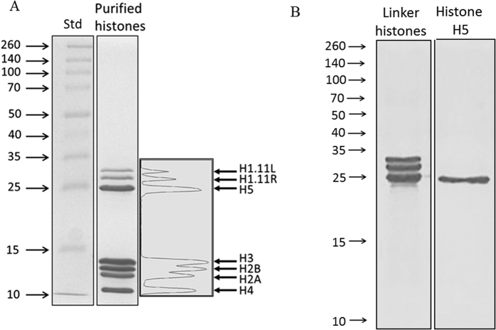
Figure 1. SDS-PAGE analysis of the TCA-precipitated histone mixture purified from chicken erythrocytes.
(A) 15% acrylamide gel revealing 7 distinct bands which were analyzed using densitometry, and then excised for proteomics LC/MS/MS analysis. All core and linker histones were present: H1.11L, H1.11R, H2A, H2B, H3, H4 and H5 (see Table 1 for proteomics and densitometry results). (B) Linker histones and histone H5 separated by SDS-PAGE on a 15% gel. Lane (1) linker histones; lane (2) purified histone H5.

