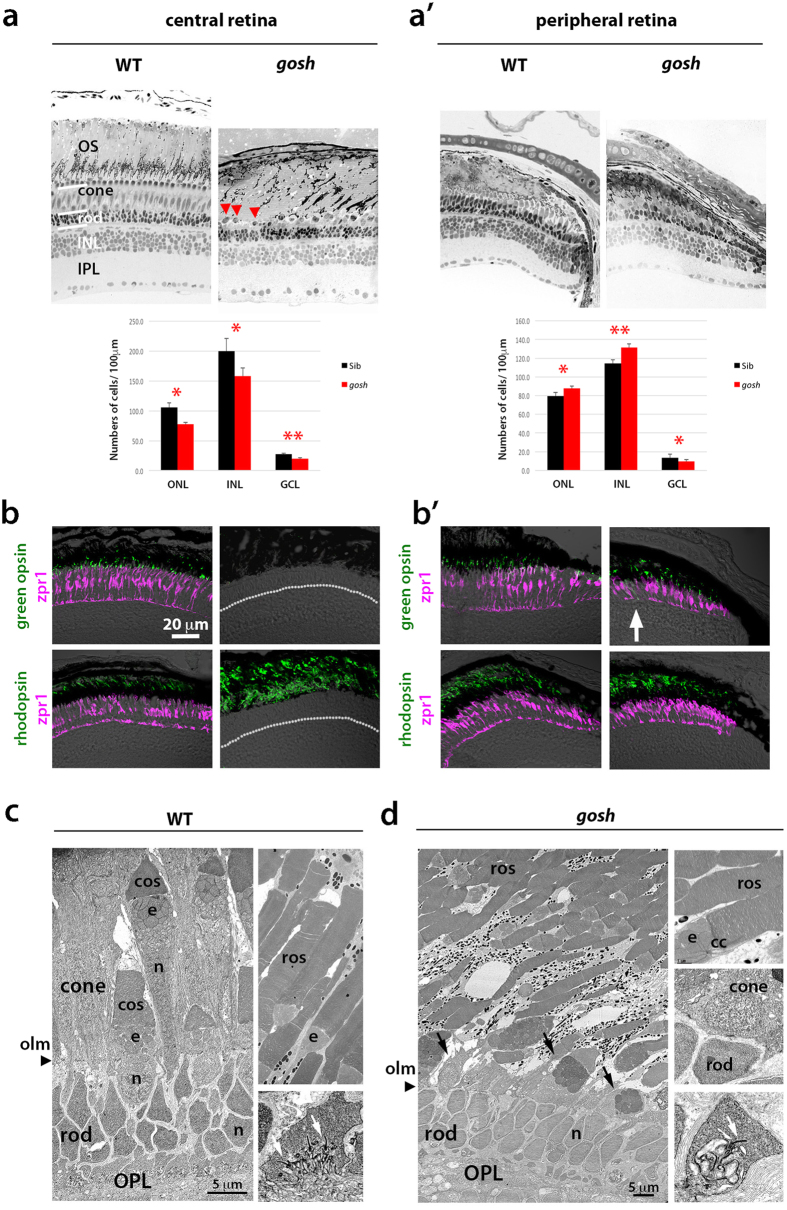
Figure 2. Cones are progressively eliminated in gosh mutants at 12 wpf.
(a) (Upper) Sections of 12 wpf wild-type and gosh mutant retinas. In wild-type central retinas, rods and cones are distinguished. In gosh mutant central retinas, most ONL nuclei have a rod-like shapes, except for a single row of short single cones (red arrowheads), and an enlarged OS area. (a’) In the gosh mutant CMZ, both rods and cones are present. (Lower) Cell number of the ONL, INL, and GCL at 12 wpf in wild type (black) and the gosh mutant (red). Numbers of nuclei within 100 μm length for each layer were counted (n = 3). The ONL, INL, and GCL cells were reduced in the central retina of the gosh mutant. In the peripheral retina, ONL and INL cells was increased, but GCL cells were decreased in the gosh mutant. Students’ t-test: *p < 0.05; **p < 0.01. (b) Labeling of 12 wpf wild-type and gosh mutant retinas with anti-green opsin and rhodopsin antibodies (green), and zpr1 antibody (magenta). In wild-type central retinas, green opsin and rhodopsin are located in cone and rod OSs, respectively. However, in the gosh mutant, green opsin is absent, while most of the OS is labeled with rhodopsin. (b’) In the gosh mutant CMZ, both green opsin and rhodopsin are detected, although green opsin is mislocalized in some cells (arrows). A dotted line indicates the interface between the ONL and the OPL. (c) Twelve wpf wild-type ONL, which consists of small, compact rod nuclei, short and long single cones, and double cones (left), rod OS (right upper), and synapses (arrows show ribbon, right bottom). An arrowhead indicates the outer limiting membrane. (d) Twelve wpf gosh mutant ONL, which consists of rod nuclei and single row of cone-like cells (arrows, left), rod OS (right upper), and synapses (a white arrow shows the synaptic ribbon, right bottom). The outer limiting membrane is maintained at the outer border of rod nuclei (arrowhead). OS, the outer segment; INL, inner nuclear layer; IPL, inner plexiform layer; OPL, outer plexiform layer; olm, outer limiting membrane; ros, rod outer segment; cos, cone outer segment; e, ellipsoid; cc, connecting cilium.

