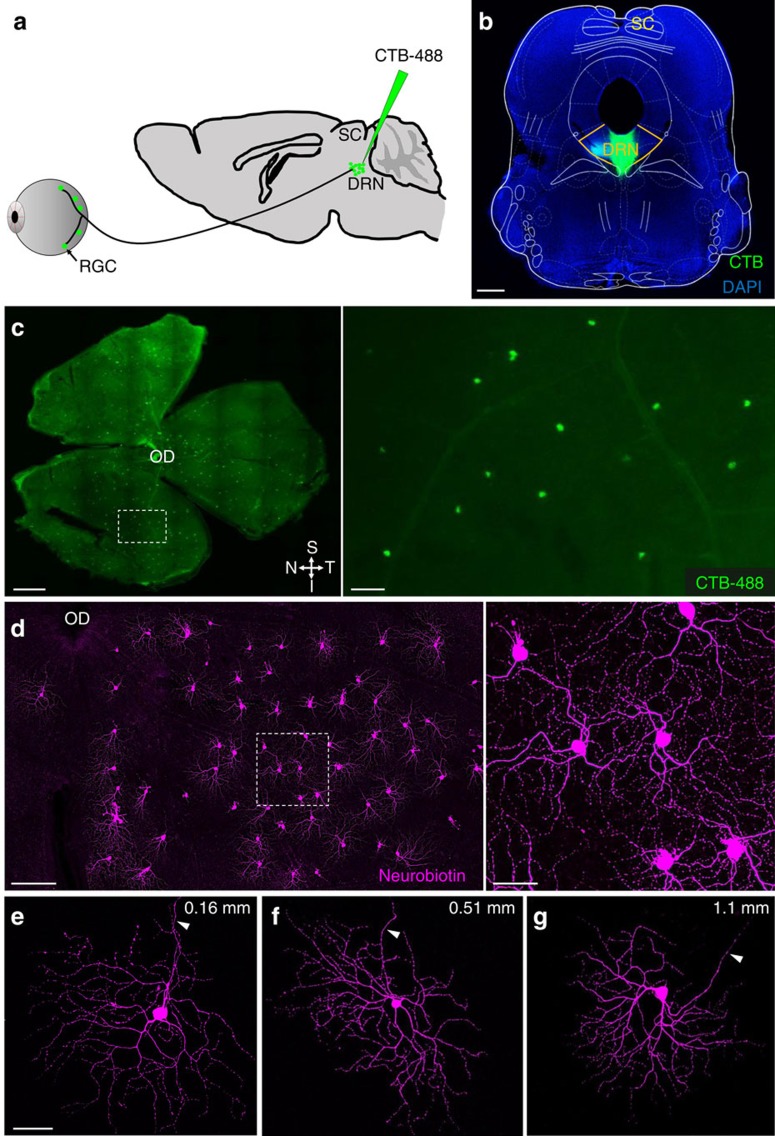
Figure 1. Retinal ganglion cells (RGCs) innervate the mouse DRN.
(a) Angled trajectory used to eject cholera toxin B subunit Alexa Fluor 488 (CTB-488) into the dorsal raphe nucleus (DRN) while avoiding the superior colliculus (SC). (b) Image of a CTB-488 injection site in the DRN. Note absence of CTB-488 in the SC; yellow lines demarcate borders of the DRN. (c) Left: retinal whole mount illustrating retrogradely labelled DRN-projecting RGCs. Retina orientation: S=superior, I=inferior, N=nasal and T=temporal. Right: region defined in white box viewed under higher magnification. (d) Left: intracellularly filled DRN-projecting RGCs in a retinal whole mount. Right: region defined in white box viewed under higher magnification. (e–g) Intracellularly filled DRN-projecting RGCs located at different retinal eccentricities relative to optic disc (OD). Arrowheads point to axons. Numbers in the upper right corner indicate distance between soma and optic disc. Scale bars: (b,c-left) 500 μm; (c-right, d-right and e) 50 μm; (d-left) 100 μm.

