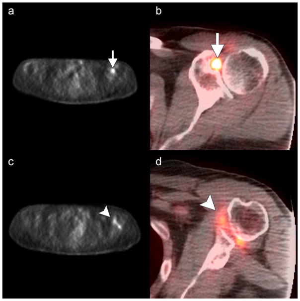
Fig. 1.
Focal uptake at the rotator interval and axillary pouch. Axial unfused (a and c) and fused (b and d) 18F-FDG PET/CT images in a 64-year-old man with mantle cell lymphoma reveals focal increased uptake at the a–b) rotator interval (arrows, SUV=5.8) and c–d) inferior capsule (arrowheads, SUV=3.5).

