
Figure 9.
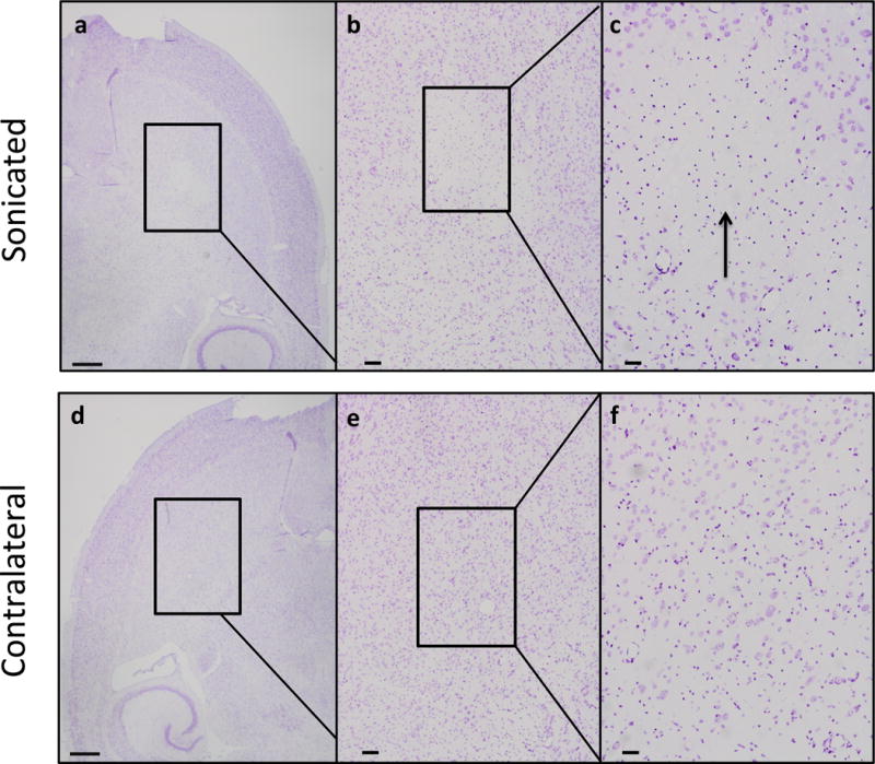
Nissl staining of paraffin embedded sections for the positive control (1.5 MPa) sacrificed 24 hours post sonication showed a loss of neurons (a–c). Other neuronal bodies, however, remained intact. For the contralateral hemisphere of the same subject, there was no pervasive abnormal morphology (d–f). Brains were sectioned horizontally at 6 μm. Scale bars: a, 1mm; b, 200 μm; c, 50 μm.