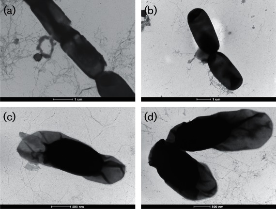
Fig. 4.

Transmission electron microscopy (negative staining with 2 % uranyl acetate) images of B. wiedmannii sp. nov. FSL W8-0169T. Panels (a) and (b) show vegetative cells, while (c) and (d) show spores with exosporium. Bars, 1 µm (a, b); 500 nm (c, d).
