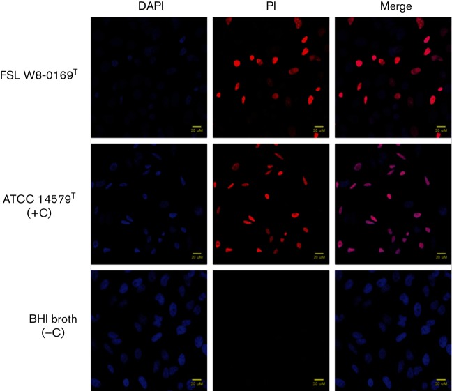
Fig. 5.
Cytotoxicity of B. wiedmannii sp. nov. FSL W8-0169T supernatants. HeLa cells grown on coverslips were co-incubated with 5 % (v/v) culture supernatants of FSL W8-0169T and B. cereus ATCC 14579T (included as a positive control), for 30min with 5 µg PI ml−1 (red). After fixation with 4 % PFA, HeLa cells were stained with 1 µg DAPI ml−1 (blue). All cells stained blue with DAPI, while only cells with compromised membrane integrity stained red. Incubation with BHI media was included as a negative control. The merged images demonstrate the proportion of cells with damaged membrane due to cytotoxic activity of bacterial supernatant.

