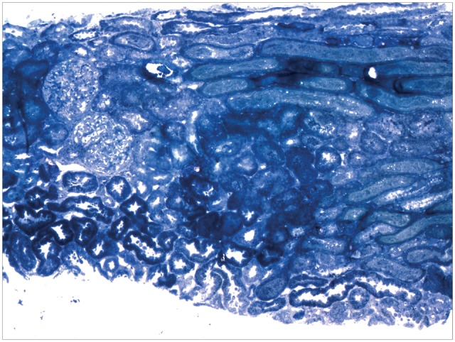
Fig. 1.

Light microscopy. Cortical zone showing two glomeruli (on left) with normal architecture and no evidence of proliferative, inflammatory or sclerosing lesions. There is no interstitial inflammation and tubules do not reveal signs of injury or necrosis (Toluidine Blue stain, original magnification ×100).
