Figure 4.
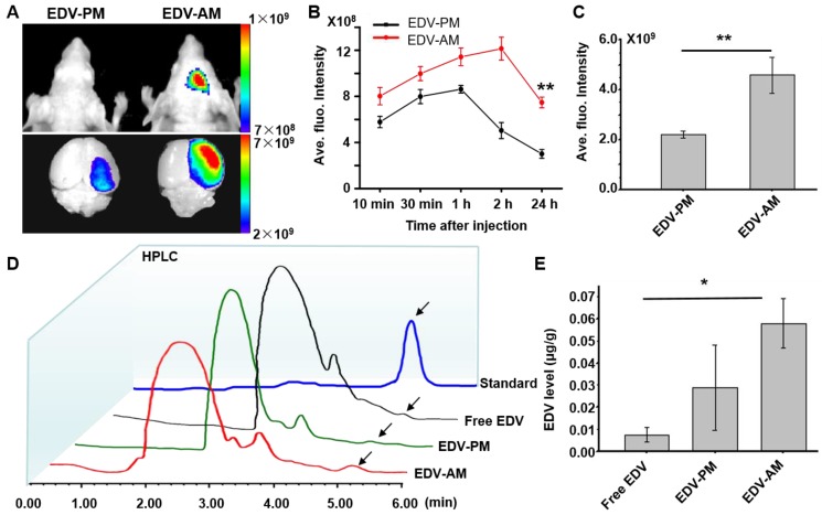
Agonistic micelles increase EDV uptake in brain ischemia. (A) NIR fluorescence images of the mouse brain at 24 h PI of EDV-AM or EDV-PM (0.078 mg EDV/mouse) via i.v.. The treatments were conducted at 6 h post-stroke model establishment. Scale unit: p/sec/cm2/Sr. (B) In vivo time-dependent normalized NIR fluorescence intensities in mouse brain area after injection of EDV-AM or EDV-PM (mean ± SD, n = 5). ** P < 0.01, EDV-AM vs. EDV-PM group. (C) Ex vivo NIR fluorescence intensities in the excised brain ischemia at 24 h PI of EDV-AM or EDV-PM (n = 5). (D) HPLC chromatography of homogenized extractions of the ipsilateral hemisphere at 24 h PI of free EDV, EDV-PM, or AM-EDV. Arrows point to the eluting peak of EDV. (E) Quantification of EDV concentration in the ipsilateral hemisphere at 24 h PI free EDV, EDV-PM or EDV-AM. * P < 0.05 EDV-AM vs. free EDV group. PI: post-injection.

