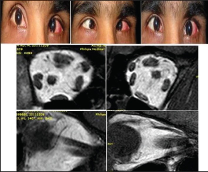
Figure 3.

Clinical picture of Case 9 showing esodeviation in both eyes in primary gaze, and limitation of abduction in both eyes. Magnetic resonance imaging showing right eye lateral rectus splitting, in both coronal section and sagittal section (left) seen in comparison to normal lateral rectus in control in coronal and sagittal section (right)
