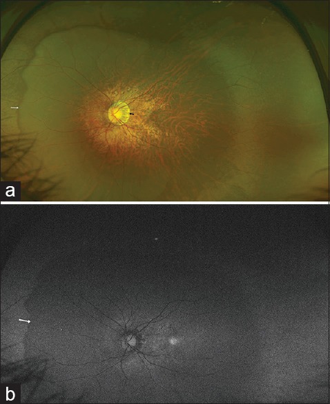
Figure 1.

(a) A large myopic conus is seen temporal to the disc (black arrow). A large posterior staphyloma is also evident. The nasal edge of the staphyloma appears as a dark crescent (white arrow). (b) An Optos autofluorescence image of the fundus showing a difference in the autofluorescence signal between the area of the staphyloma and the rest of the peripheral fundus. The white arrow marks the boundary of the staphyloma
