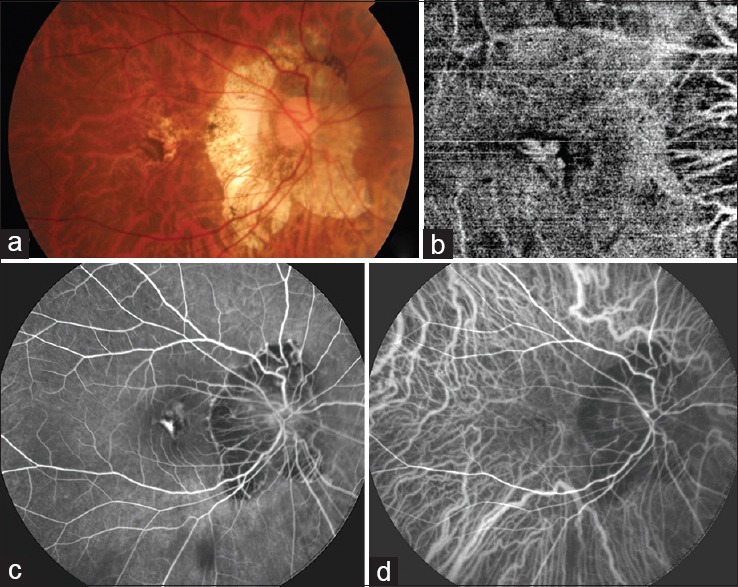
Figure 4.

(a) Fundus image of a myopic patient showing a grayish lesion at the fovea with pigmented margins, suggestive of a choroidal neovascular membrane. (b) Optical coherence tomography angiography image of the same patient showing the branching network of vessels of the choroidal neovascular membrane with a surrounding dark halo. (c) Fluorescein angiography showing the hyperfluorescent choroidal neovascular membrane. (d) Indocyanine green angiography also showing the hyperfluorescent choroidal neovascular membrane
