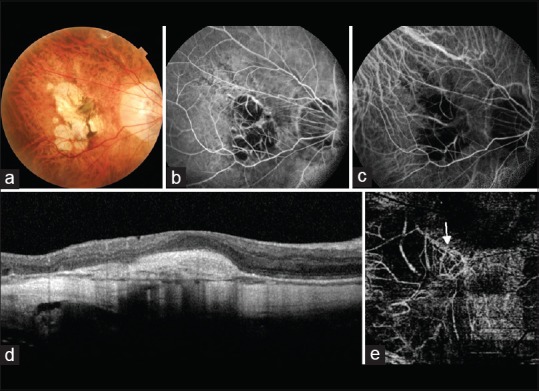
Figure 5.

(a) Fundus image of a myopic patient showing a grayish membrane at the fovea clinically suggestive of a choroidal neovascular membrane. (b) The gray membrane appears hyperfluorescent of fundus fluorescein angiography. However, the hyperfluorescence is not as marked as is seen in classic choroidal neovascular membranes. (c) Indocyanine green angiography fails to detect the choroidal neovascular membrane. (d) Optical coherence tomography shows a spindle-shaped hyper-reflective area above the retinal pigment epithelium suggestive of a choroidal neovascular membrane. There is absence of associated subretinal or intraretinal fluid. (e) An abnormal branching network of vessels (white arrow) is visible on the optical coherence tomography angiography image segmented at the deep retinal level. This is suggestive of a Type 2 choroidal neovascular membrane
