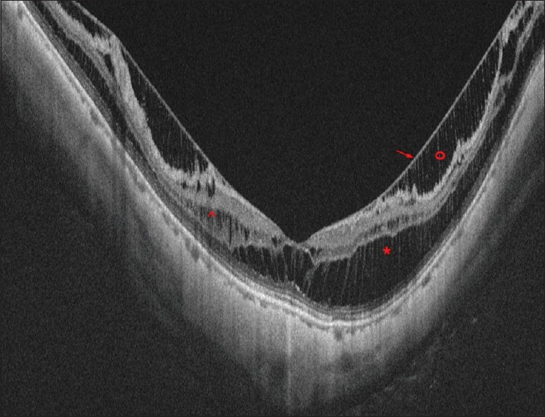
Figure 6.

Optical coherence tomography image of a case of myopic maculoschisis with posterior staphyloma. Red * shows the schisis in the outer plexiform layer. Red ^ shows the schisis in the inner nuclear layer. Red o shows the schisis in the nerve fibre layer and the red arrow shows the overlying stretched internal limiting membrane
