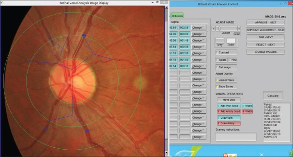
Figure 1.
Window interface of retinal vessel diameter measurement software (IVAN). The fundus is divided into four areas (the optic disc zone, A zone, B zone, and the perimeter zone from inside to outside) by three rings (disc diameter ring, 1/2 optic disc diameter away from the edge of the optic disc ring, and 1 optic disc diameter away from the edge of the optic disc ring from inside to outside), and the diameter of artery and vein was measured in different regions.

