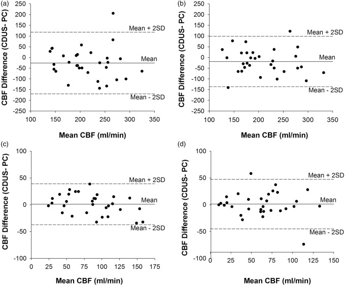
Figure 4.
Bland–Altman plots of differences in CBF measurements between CDUS and PC-MRI. Left: internal carotid artery (a); right: internal carotid artery (b); left: vertebral artery (c); and right: vertebral artery (d).
CBF: cerebral blood flow; CDUS: color-coded duplex ultrasonography; PC-MRI: phase contrast magnetic resonance imaging.

