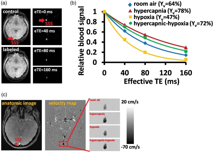
Figure 2.
Representative MR images for the CMRO2 measurement. (a) TRUST MRI for the measurement of global Yv. Red arrow indicates the SSS. The subtraction of control and labeled images yielded pure blood signal. (b) The fitting of the signal as a function of effective TE can generate an estimation of blood T2. Data from four different respiratory states are shown. (c) Phase contrast MRI for the measurement of CBF. Darker color indicates a greater flow velocity.
CMRO2: cerebral metabolic rate of oxygen; TRUST : T2-Relaxation-Under-Spin Tagging; SSS: superior sagittal sinus.

