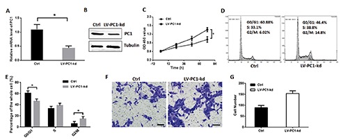
Figure 3.

PC1 knockdown promoted VSMC proliferation. A) VSMC was infected by LV-PC1-kd for 72 h, and Q-PCR was used to detect the efficiency. B) Western blot was used to detect the efficiency of LV-PC1-kd. C) Cell proliferation was detected through CCK8 kit and valuated by an absorbance at 450 nm; two groups of VSMCs were detected: one served as control, and the other was infected with lentivirus containing PC1-shRNA that can knockdown PC1. D) Cell cycle of two groups of VSMCs detected through flow cytometry in a representative experiment. E) Cell ratios in the G0/G1, S, and G2/M phases of the two VSMC groups. F) Transwell assay of PC1-kd VSMCs in a representative experiment. The VSMCs migrated through transwell chamber were colored purple by 0.05% crystal violet; scale bars: 50 μm. G) The number of migration cells in the transwell assay of each VSMC group. Each experimental condition was performed in triplicate and applied in three independent experiments. Values are expressed as mean ± SD; *P<0.01.
