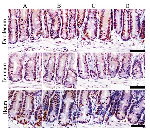
Figure 6.

The changes of PCNA-positive immunity reaction cells in intestine of mice (immunohistochemistry staining). The reaction cells appear brown in colour and are mostly located in the base of intestine villi and intestinal crypts. A) Control group. B) Stress-restraint group. C) CH-treated group. D) stress-restraint + CH group. Scale bars: 50 µm.
