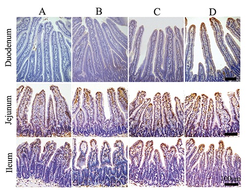
Figure 8.

The distribution and morphology of apoptotic cells in intestine of the different treatment mice (TUNEL). The apoptotic cells appear brown in colour, and apoptotic epithelial cells were found predominantly in the tip extrusion zone of villus; A) Control group. B) Stress-restraint group. C) CH-treated group. D) stress-restraint + CH group. Scale bars: 100 µm.
