Figure 1.
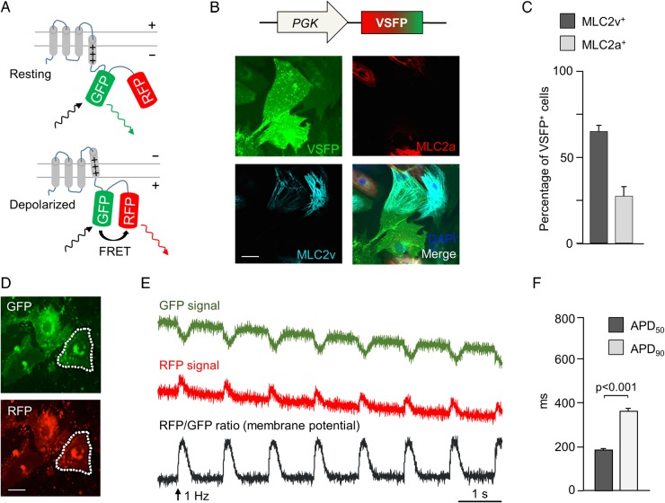
Förster resonance energy transfer-based optical membrane potential recordings in human-induced pluripotent stem cells-derived cardiomyocytes. (A) Mode of action of the membrane potential sensor voltage-sensitive fluorescent protein. A voltage-sensing transmembrane protein is linked to a pair of a green and a red fluorescent protein. Upon depolarization, GFP and RFP are brought closer together, increasing Förster resonance energy transfer, which makes GFP appear dimmer and RFP brighter. (B) Pseudocolour images of human-induced pluripotent stem cells-derived cardiomyocytes infected with PGK-voltage-sensitive fluorescent protein lentivirus and stained for MLC2v (cyan) and MLC2a (red). (C) Percentage of cells expressing MLC2v and MLC2a among voltage-sensitive fluorescent protein-expressing cells (n = 325 cells). (D) GFP and RFP pseudocolour images of voltage-sensitive fluorescent protein-expressing human-induced pluripotent stem cells-derived cardiomyocytes (dotted lines: region of interest used to quantify fluorescence signal). (E) Background-corrected GFP and RFP fluorescence signals recorded at 100 Hz upon field stimulation at 1 Hz. An RFP/GFP ratio was calculated as the membrane potential signal. (F) Action potential duration (APD50 and APD90) in PGK-voltage-sensitive fluorescent protein-expressing cells. N = 92 cells.

