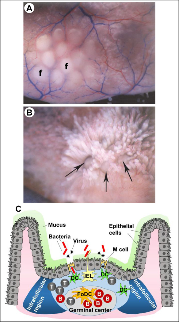
Figure 2.
Peyer’s patches (PPs). (A) A PP consisting of 12 lymphoid follicles (f) as seen in a gerbil intestine. (B) Lymphoid follicles in a gerbil intestine lined by intestinal villi. The large arrow points to the lymphoid follicle dome. (C) Schematic of a PP showing M cells and the different immune cell populations. Note the absence of mucus on top of M cells.
T: T cells, B: B cells, DC: dendritic cells, IEL: intraepithelial lymphocyte, FoDC: follicular dendritic cell, IFR: intra-follicular region. Figures 2A and 2B adapted with permission from publisher of [23]

