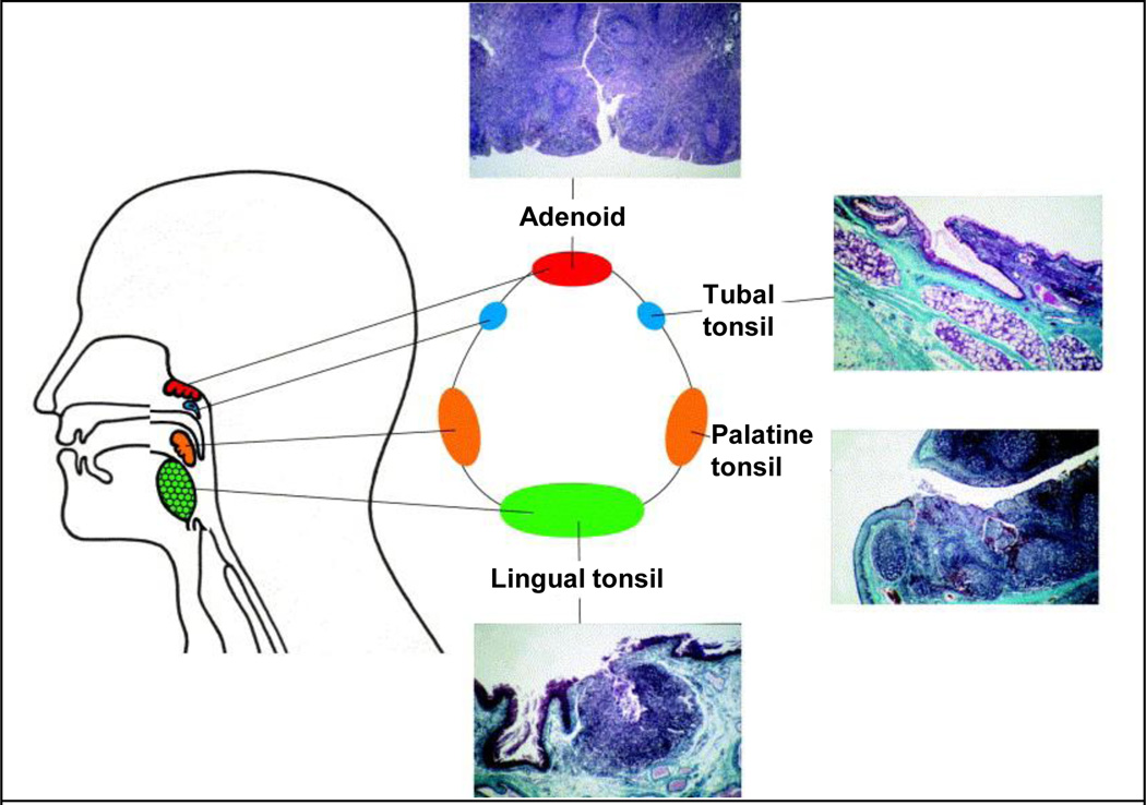
Figure 3.
Lymphoid tissues of the Waldeyer’s ring and their histological sections. Sections show crypts (white indentations in histology sections) that increase surface area available for direct antigenic uptake and stimulation. Adapted with permission from publisher of [28]

