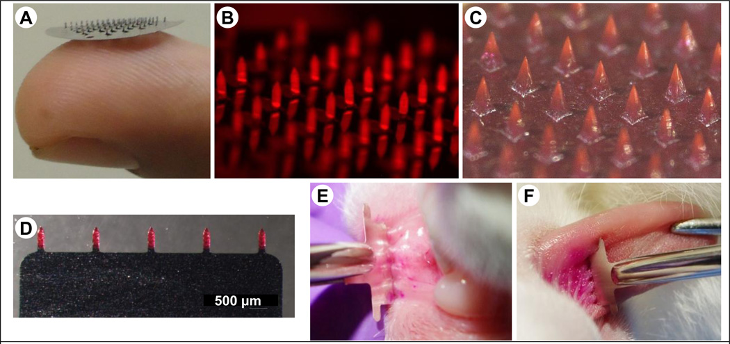
Figure 7.
Microneedle devices. (A) Photograph of a stainless steel microneedle patch resting on a fingertip, (B) Stereomicrograph of a stainless microneedle patch with microneedles coated with fluorescent ovalbumin, (C) Stereomicrograph of a polymer microneedle patch (image courtesy of Georgia Institute of Technology, (D) Stereomicrograph of a planar stainless steel microneedle patch coated with sulforhodamine, (E) A planar microneedle patch being inserted into the inner surface of the lower lip of a rabbit, (F) A planar microneedle patch being inserted into the dorsal surface of a rabbit tongue.

