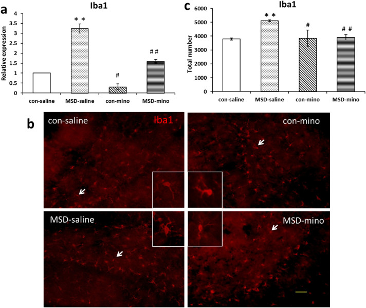
Figure 3. Minocycline treatment reduced microglia activation caused by MSD in hippocampus of young offspring rats.
(a) Relative mRNA expression of Iba1 was increased after MSD, and the expression of Iba1 was decreased by minocycline treatment. (b) Representative example of microglia and its morphology in the DG of hippocampus. (c) The total number of Iba1+ cells also increased in the MSD-saline offspring rats, the microglia show large somas, short thick processes and a rounded amoeboid morphology, while the number decreased after minocycline injections and the microglial morphology returned to resting state. The arrows indicate typical microglia. * P < 0.05, ** P < 0.01 vs. the control-saline. # P < 0.05, # # P < 0.01 vs. the MSD-saline. Values are the mean ± SEM. Scale bars: 10 μm.

