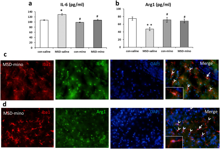
Figure 6.
Minocycline altered IL-6 and Arg1 protein expression derived from activated microglia in the hippocampus of young offspring rats Hippocampal IL-6 protein level increased (a) and Arg1 (b) protein level decreased in the MSD-saline offspring. These changes were restored to the level of control-saline rats after minocycline treatment. Representative fluorescent images of the MSD-mino brain slices examined for Iba1 and IL-6 (c), Iba1 and Arg1 (d) immunoreactivity in the DG of hippocampus in the young offspring rats. The color of microglia is red, IL-6 and Arg1 are green. Arrowheads represent activated microglia, which released IL-6 (c) or Arg1 (d), arrows indicate fluorescent images of Iba1 and IL-6 or Arg1. * P < 0.05, ** P < 0.01 vs. control-saline. # P < 0.05 vs. MSD-saline. Values are the mean ± SEM. Scale bars: 20 μm.

